Human T Lymphocyte Photograph by Dennis Kunkel Microscopy/science Photo
Multiplex Fluorescence Microscopy With Four Lymphocyte Cell Markers: CD3, CD4, CD8, CD20, and CD68 as Reference. All lymphocyte markers were detected within the 1 mm 2 ROIs around mesh fibers that marked the FBG, but CD20+ cells were mainly seen in clusters outside the FBG (Figure 4). In close vicinity to the fibers, there were predominantly.
Reactive Lymphocyte 1 ERA
Find Lymphocyte microscope stock images in HD and millions of other royalty-free stock photos, illustrations and vectors in the Shutterstock collection. Thousands of new, high-quality pictures added every day.

Lymphocyte cells stock photo. Image of care, microscopic 92594228
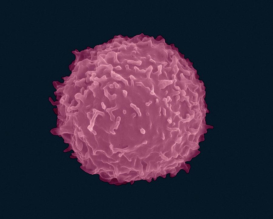
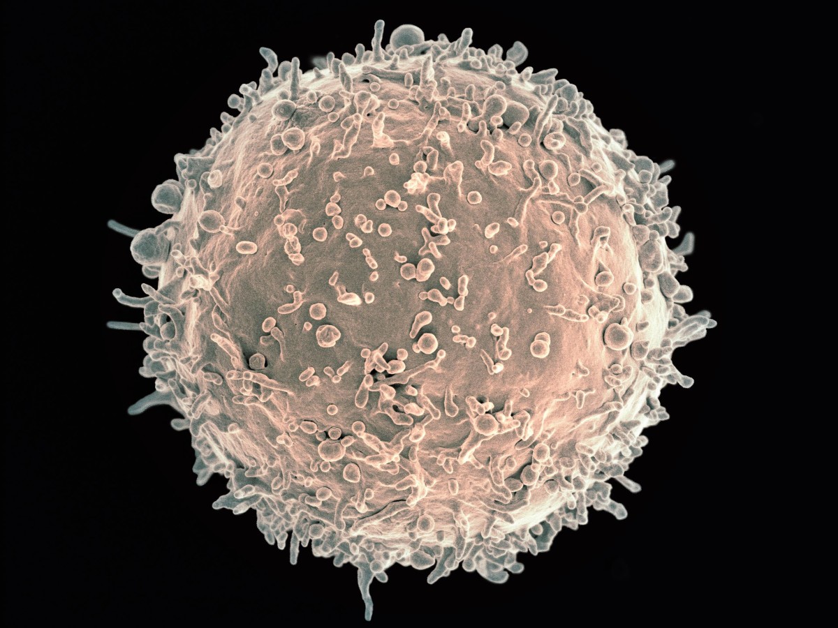
Lymphocyte: A scanning electron microscope (SEM) image of a single human lymphocyte. B cells are involved in humoral adaptive immunity, producing the antibodies that circulate through the plasma. They are produced and mature in bone marrow tissues and contain B cell receptors (BCRs) that bind to antigens. While in the bone marrow, B cells are.

Autoimmune diseases may be side effect of a strong immune system New
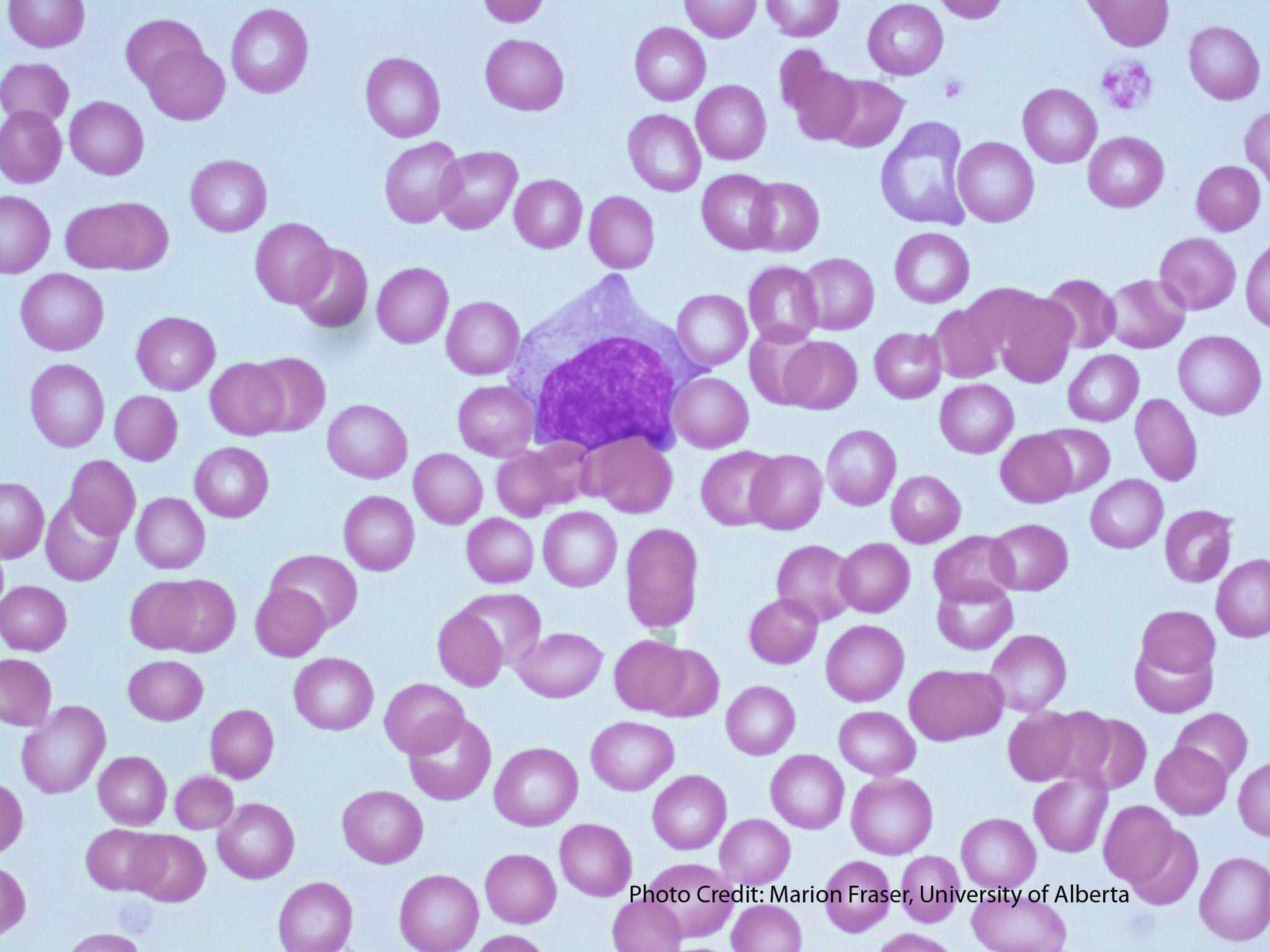
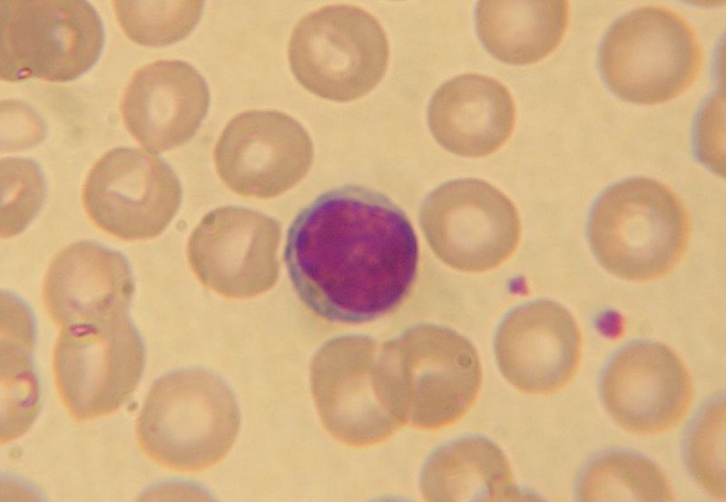
Microscopic appearance of the blood. Lymphocyte Lymphocytus 1/6 Synonyms: none Lymphocytes are types of leukocytes (white blood cells) that represent the primary cells of the immune system. All leukocytes are classified into granulocytes and agranulocytes, and lymphocytes belong to the agranulocyte group.
The Immune System, Lymphocytes, and NK, B, and T Cells Owlcation
The validation set contained two images of each considered organ (breast, colon, prostate), one stained with CD3 and one stained with CD8. The test set contained fifteen images of colon cancer and breast cancer, and ten images of prostate cancer, with the same proportion of slides stained with CD3 and CD8. 3. Learning to detect lymphocytes
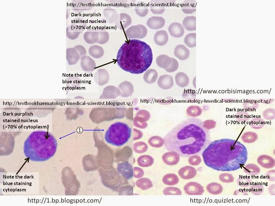
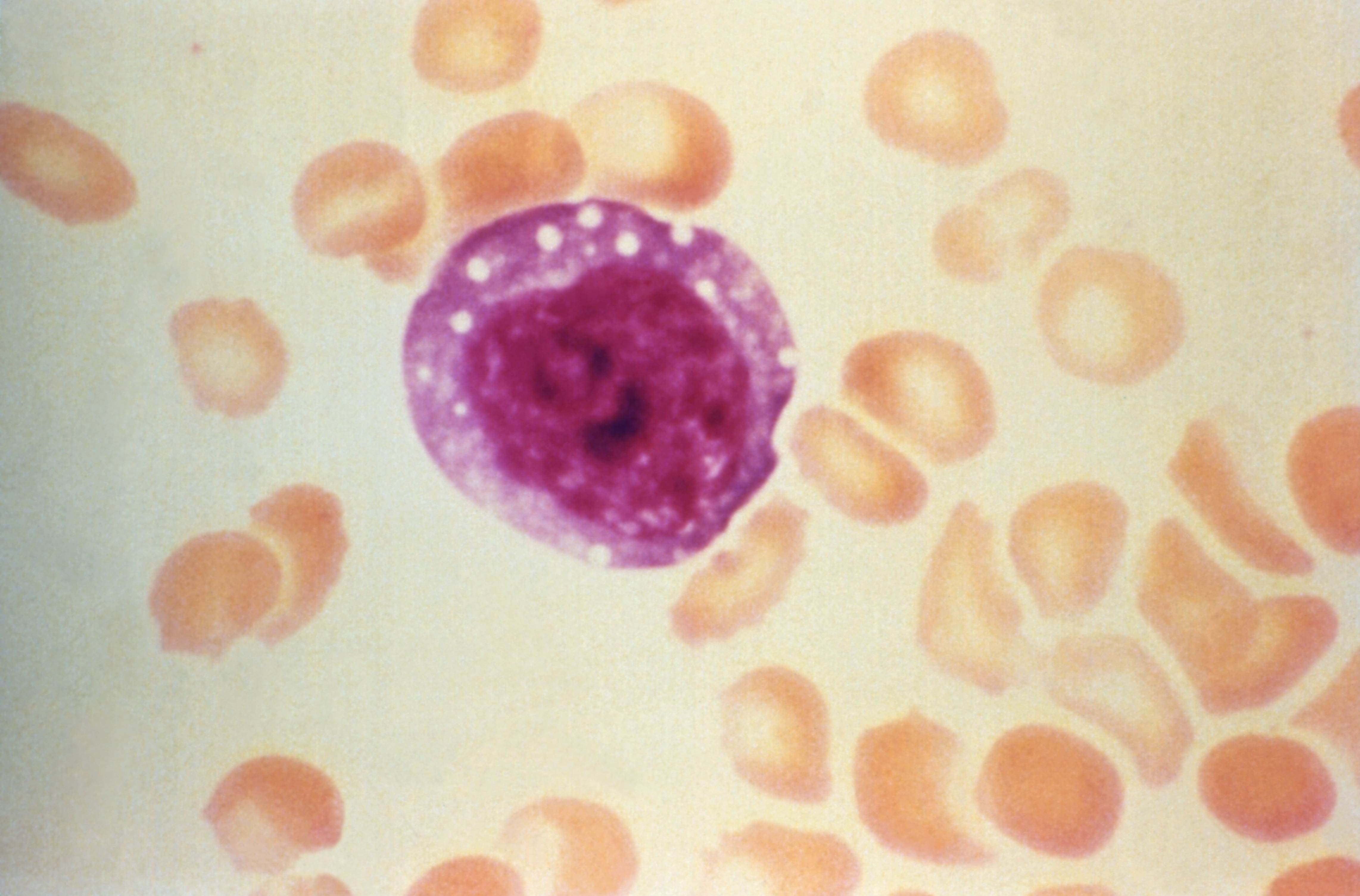
Haematology in a NutShell Reactive/Atypical Lymphocytes
Lymphocytes are a central component of immune defence mechanisms and have a pivotal role in our battle against pathogens. During adaptive immune responses, lymphocytes bearing antigen receptors.
Wright's stain wikidoc
Lymphocyte Microscopy To identify these cells in a blood smear, Wright's stain can be used. This is an important stain that is often recommended for differential staining of blood smears or bone marrow. Requirements Blood sample immediately drawn or stored in an EDTA tube. Glass slides (2 or 3) Cover slip Compound microscope Wright's stain
Free picture micrograph, atypical, enlarged, lymphocyte, blood smear
To bridge this gap between in vivo and in vitro approaches, we used two-photon microscopy ( 5, 6) to image individual living T and B lymphocytes deep within the intact lymph node. Purified T and B cells from donor BALB/c mice were labeled with green [5- (and 6-) carboxyfluorescein diacetate succininyl ester (CFSE)] or red (5- (and-6)- ( ( (4.
Large granular lymphocyte in a patient of systemic lupus
The proposed method was tested on live lymphocyte images acquired through the phase-contrast microscope from the blood samples of mice, and comparative experimental results showed the advantages of the proposed method in terms of the accuracy and the speed. Tracking experiments showed that the proposed method can accurately segment and track.
Clefted Lymphocyte 1.
Human lymphocyte microscope Stock Photos and Images (269) See human lymphocyte microscope stock video clips Quick filters: Cut Outs | Vectors | Black & white Sort by Relevant RM 2DF79FR - Human lymph node or lymph gland. Photomicrograph. RM 2JKFT4D - Scanning electron micrograph of a human natural killer cell. Credit: NIAID
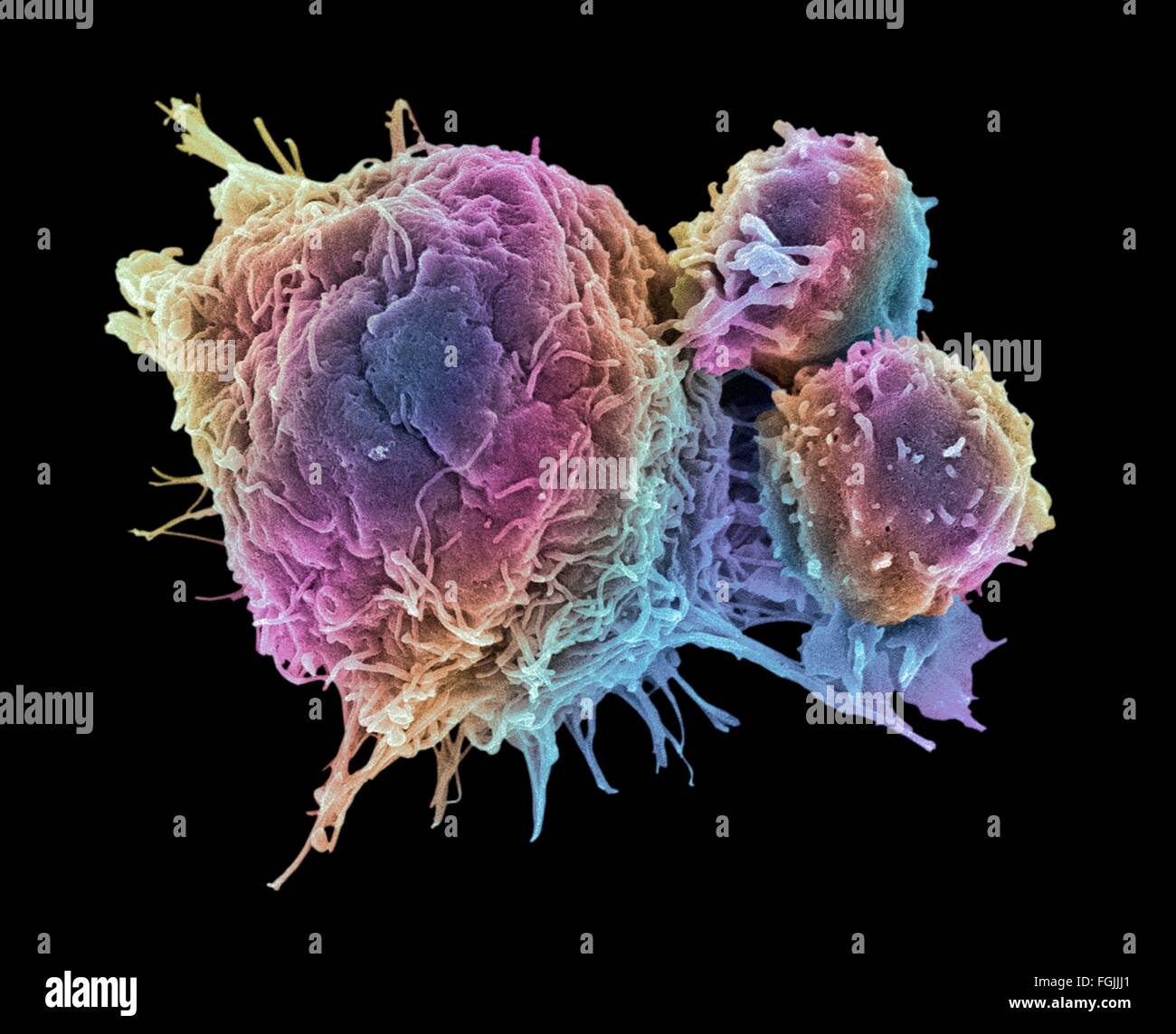
T lymphocytes and cancer cell. Coloured scanning electron micrograph
In this case, we perform a two-dimensional (2D) tiled scan of a 16 mm × 6 mm area using inverted epifluorescent microscopy while passing the tiled images to the YOLOv5 network (Fig. 2a). For a.

Lymphocytes bas, élevés définition, causes et examen
[2] Types A stained lymphocyte surrounded by red blood cells viewed using a light microscope 4D live imaging of T cell nuclear dynamics viewed using holotomography microscopy Giemsa stained lymphocytes in peripheral blood The three major types of lymphocyte are T cells, B cells and natural killer (NK) cells. [2]

Infectious mononucleosis and atypical lymphocytosis on a smear
Time-lapse imaging provides a uniquely dynamic view of biological processes in living systems 1,2,3,4,5,6,7. Powerful insights into development and function have followed through a union of modern.

Pin on medical education
Intravital confocal microscopy and two-photon microscopy are powerful tools to explore the dynamic behavior of immune cells in mouse lymph nodes (LNs), with penetration depth of ~100 and ~300.
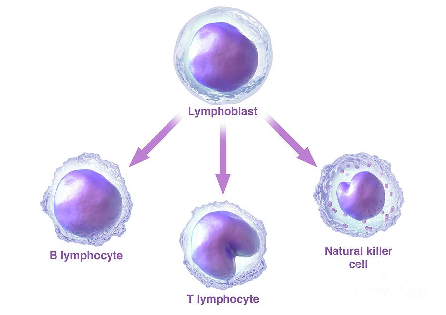
Lymphocyte Formation From Lymphoblasts Photograph by Maurizio De
In particular, when combined with two-photon laser microscopy, intravital imaging of surgically exposed lymph nodes provides a unique view of lymphocyte migration and antigen presentation as it occurs within the living animal.

Large Lymphocyte
Find Lymphocyte T Microscope stock images in HD and millions of other royalty-free stock photos, illustrations and vectors in the Shutterstock collection. Thousands of new, high-quality pictures added every day.